Figure 2.

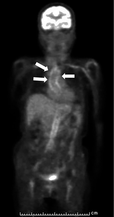
Coronal section of 18F-FDG PET/CT. Abnormal FDG uptakes were observed in the patient’s aortic wall and aortic branches (white arrow).

Coronal section of 18F-FDG PET/CT. Abnormal FDG uptakes were observed in the patient’s aortic wall and aortic branches (white arrow).