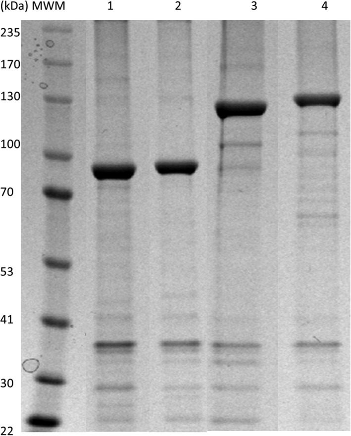
Figure 1.

SDS–PAGE analysis of proteins attached to various polyester beads isolated from Clear coli. Lane 1, Cpe30‐Rv1626 beads (92.5 kDa); lane 2, CS.T3‐Rv1626 beads (93.1 kDa); lane 3, Fla66‐Rv1626 beads (134 kDa); lane 4, Cpe30‐CS.T3‐Fla66‐Rv1626 beads (141 kDa). Corresponding genes were inserted into pPOLYC‐Rv1626 plasmid using XhoI/BsrGI sites. The linker VLAVAIDKRGGGGG (hydrophobic‐charged amino acids) is included in this plasmid between PhaC and Rv1626 to facilitate display of the fusion partners (Jahns and Rehm, 2009). Proteins were quantified by SDS–PAGE gel densitometry.
