Figure 2.

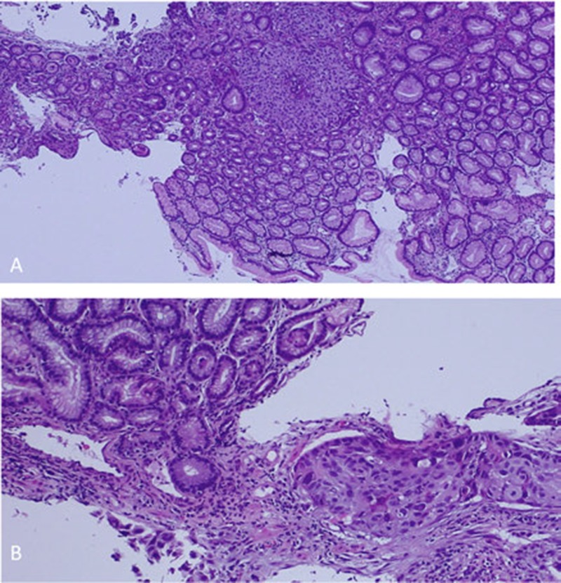
Histopathological examination: hematoxylin and eosin stain showing invasive squamous cell carcinoma under uninvolved gastric mucosa. Low (A) and High (B) magnification.

Histopathological examination: hematoxylin and eosin stain showing invasive squamous cell carcinoma under uninvolved gastric mucosa. Low (A) and High (B) magnification.