Figure 1.
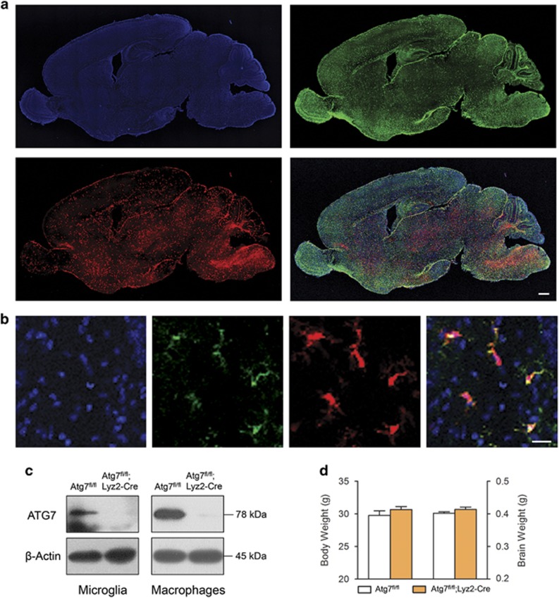
Phenotypes of mice with Atg7-deficient microglia. (a) Sagittal images of Atg7fl/+;Lyz2Cre/+;ROSA26-tdTomato mouse brain at postnatal day 7 stained with an antibody against Iba-1 (green) and Hoechst 33342 (blue). Tomato expression (red) was detected in all brain regions. Scale bar=500 μm. (b) Most of the Tomato expression (red) was co-localized with Iba-1-positive microglia (green). Scale bar=20 μm. (c) Atg7 protein was not detected in the lysate of primary microglia cultures or in peritoneal macrophages collected from Atg7fl/fl;Lyz2-Cre mouse. (d) Body and brain weights of adult Atg7fl/fl;Lyz2-Cre mice were similar to those of controls (n=12 for each group).

