Figure 4.
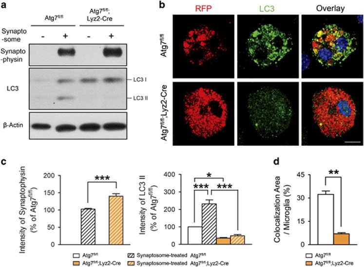
Impaired degradation of the synaptosome by atg7-deficient microglia. (a, c) Primary microglia from Atg7fl/fl or Atg7fl/fl;Lyz2-Cre mice were cultured and isolated synaptosomes from red fluorescent protein (RFP)-expressing mouse brain were added to the microglial cultures. Western blots showed that synaptophysin is increased and LC3-II is decreased in atg7-deficient microglia compared with wild-type microglia. One-way analysis of variance with Tukey’s post hoc test. F(3,11)=355.40, P<0.001 (synaptophysin). F(3,11)=57.19, P<0.001 (LC3-II). n=3 for each group. (b, d) Immunocytochemistry of microglia showing that punctated forms of LC3 (green) were increased in wild-type microglia, with co-localization (yellow) of RFP-positive synaptosomes (red). LC3 signals were diffuse and the RFP signal was increased in atg7-deficient microglia compared with wild-type microglia. Scale bar=20 μm. *P<0.05, **P<0.01, ***P<0.001 (two-tailed Student’s t-test).

