Figure 5.
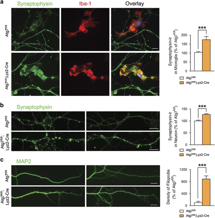
Neuronal changes after their co-culture with atg7-deficient microglia. (a) Primary mouse cortical neurons (18 days in vitro) were co-cultured with primary microglia from Atg7fl/fl or Atg7fl/fl;Lyz2-Cre mice for 2 weeks and then immunostained with antibodies against synaptophysin (green) and Iba-1 (red). Greater accumulation of synaptophysin was seen in atg7-deficient Iba-1-positive microglia than in wild-type microglia. Scale bar=20 μm. (b) Synaptophysin puncta were increased in neurons co-cultured with atg7-deficient microglia. Scale bar=20 μm. (c) MAP2-positive dendrites showed increased filopodia in neurons co-cultured with atg7-deficient microglia. Scale bar=20 μm. ***P<0.001 (two-tailed Student’s t-test).

