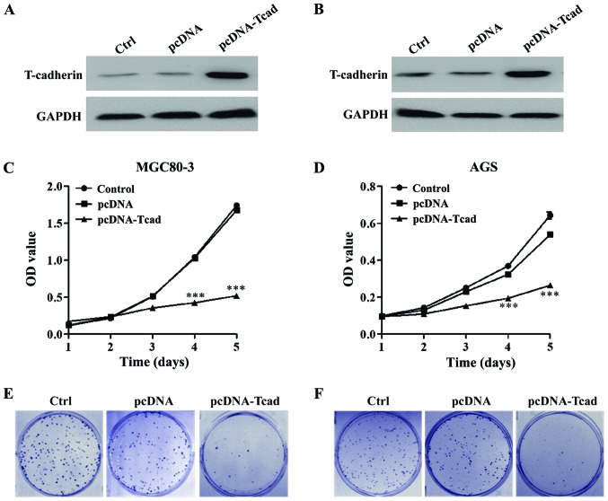
Figure 2.
T-cadherin inhibits GC cell proliferation. Overexpression of T-cadherin protein in transfected cell lines, (A) MGC80-3 and (B) AGS was confirmed by western blotting. MTT assay was used to measure cell viability in (C) MGC80-3 and (D) AGS after transfection with empty vector pcDNA or pcDNA-T-cadherin (pcDNA-Tcad). These data are shown as the mean ± standard deviation of three independent experiments. The size and number of colonies formed in (E) MGC80-3 and (F) AGS cells recorded under a light microscope. The representative pictures shown are from one of three independent experiments. ***P<0.001 vs. control or empty vector pcDNA.

