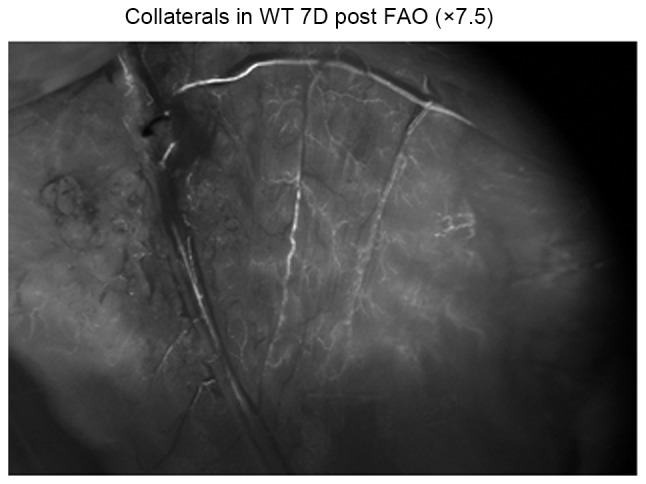
Figure 3.

Fluorescence microscopy of the collaterals with a magnification ×7.5 in wt mice 7 days post FAO. No significantly difference was observed between Gja5+/− mice and Gja5+/+ mice, the collaterals in wt mice 7 days post FAO were more than the collaterals in wt mice 3 days post FAO. LDF imaging on day 7 demonstrated that hindlimb perfusion recovered partly, compared with LDF imaging at day 3. wt, wild type; FAO, femoral artery occlusion; Gja5, gap junction protein α 5; LDF, Laser Doppler Flow.
