
Figure 1.
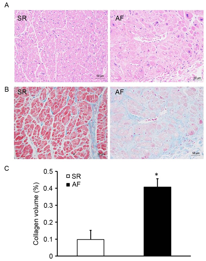
Expansive atrial myocytes and accumulated collagen in the patients with AF and rheumatic heart disease. (A) Hematoxylin and eosin staining of the right atrial tissue of patients in the SR and AF groups. (B) Masson staining of the right atrial tissue of patients in the SR and AF groups. Magnification, ×400. (C) Collagen volume fraction of the SR and AF groups. *P<0.05 vs. SR group. SR, sinus rhythm; AF, atrial fibrillation.