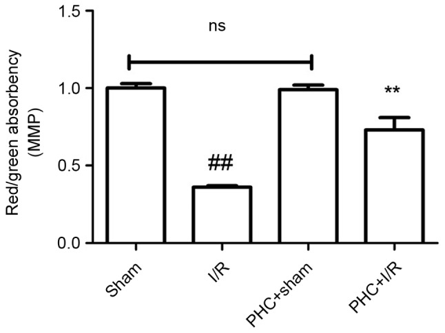
Figure 4.

Effect of PHC on the MMP. Following 3 h reperfusion, a JC-1 staining method was performed to detect MMP. The ratio of red absorbency/green absorbency represents the MMP. Data are expressed as mean ± standard deviation (n=6). ##P<0.01 vs. sham; **P<0.01 vs. I/R. ns, not significant (P>0.05); I/R, ischemia/reperfusion; PHC, penehyclidine hydrochloride; MMP, myocardial cell mitochondrial membrane potential.
