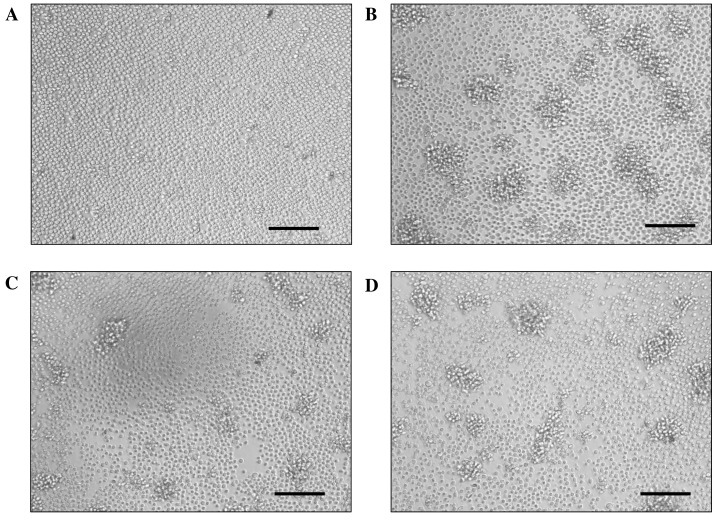
Figure 3.
Microscopic images of Con-A-stimulated mouse spleen cell cultures. Representative images of spleen cells without and with Con-A are shown in (A and B), respectively. Representative images of spleen cells stimulated with Con-A treated with WEBP (1:50 and 1:150 dilution of extract in water) are shown in (C) and (D), respectively. Scale bar,100 µm. WEBP, water extract from bell pepper leaves.

