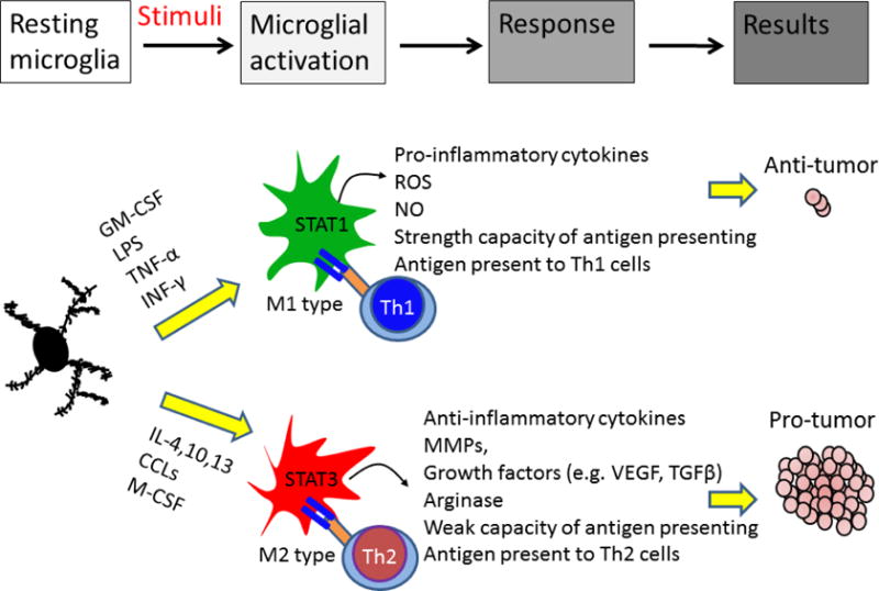
Figure 2. Differential roles of activated microglia/macrophages in the brain tumor.

Microglia/macrophages have both pro- and anti-tumor potentials. In response to granulocyte-macrophage colony stimulating factor (GM-CSF), lipopolysaccharide (LPS), tumor necrosis factor-α (TNF-α), and interferon-γ (INF-γ) stimuli, microglia/macrophage can be polarized to M1 phenotype. M1 cells exhibit anti-tumor immunity by producing cytotoxic factors and presenting tumor antigen to T helper type 1 cells (Th1) cells. STAT1 activation in M1 cells induces pro-inflammatory cytokines production and increases T-cell-mediated cytolytic activity, leading to tumor cell damage. In response to interleukin-4 (IL-4), chemokine (C-C motif) ligands (CCLs) and macrophage colony-stimulating factor (M-CSF), microglia/macrophage polarize into M2 phenotype. M2 cells express STAT3 that induces anti-inflammatory factors. M2 cells also modulate Th2 cells, which promotes tumor progression. In addition, M2 cells can promote tissue repair and angiogenesis, resulting in tumor progression.
