Fig. 1.
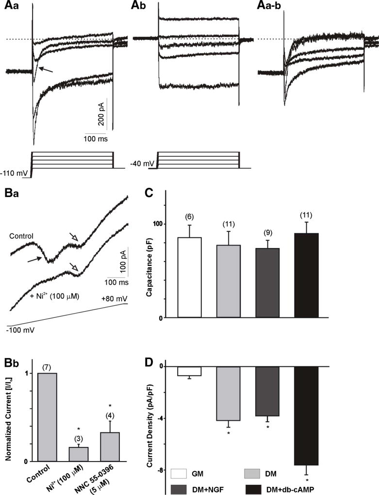
Whole cell Ca2+ currents in ND7-23 cells. Aa–b Example of whole cell Ca2+ currents generated in a differentiated ND7-23 cell. Current traces obtained from a holding potential of −110 mV (Aa) or from a holding potential of −40 mV (Ab). In this and subsequent figures, the voltage step protocol is shown below the current trace. Note that the transient component (arrow) generated by voltage steps from a holding potential of −110 mV was eliminated when the currents were generated from a holding potential of −40 mV. Aa–b Digital subtraction of Aa and Ab traces shows the inactivating component and represents the LVA (or T-type) Ca2+ currents used for further studies. B Example of whole cell Ca2+ currents generated in a differentiated ND7-23 cell by a voltage ramp before and after the application of 100 µM NiCl2. Note the two peaks generated by the activation of LVA and HVA Ca2+ channels (Ba). Only the peak current generated by activation of LVA Ca2+ channels was sensitive to blockade with NiCl2. Bb Treatment of differentiated ND7-23 cells with NiCl2 or NNC 55–0396 causes a significant reduction in the peak current generated by activation of LVA Ca2+ channels. In this and subsequent figures, error bars represent SEM and the number of cells recorded is provided above each bar. The asterisk denotes p < 0.05 vs. control. C Comparison of cell capacitance in ND7-23 cells before and after induction of cell differentiation with differentiation media supplemented with NGF or db-cAMP. ND7-23 cells were cultured for ~10 days with growth media (GM) or differentiation media (DM) supplemented with NGF (50 ng/mL) or db-cAMP (1 mM). D Mean T-type Ca2+ current densities generated in ND7-23 cells under different culture conditions. Note the robust stimulation of T-type Ca2+ current densities in ND7-23 cells cultured in differentiation media with or without NGF or db-cAMP. T-type Ca2+ current density was calculated from the peak current amplitude generated by a voltage step to −20 mV from the digital subtraction of current traces at −110 and −40 mV. The asterisk denotes p < 0.05 vs. growth media (GM)

