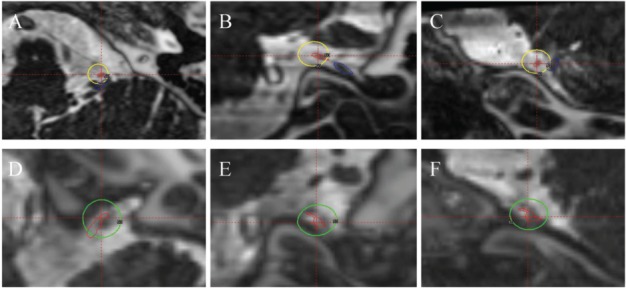
Figure 1.

MRI CISS images fused with CT from the Gamma-Plan (Elekta Instruments AB, Sweden). A, B and C) Axial, coronal and sagittal images (respectively) of the patient treated with cisternal target. The yellow outline indicates the 50% isodose line of 70 Gy dose delivered with a single 4-mm collimator. The red outlines the glossopharyngeal nerve and the blue outlines the vagus nerve. D, E and F) Axial, coronal and sagittal images (respectively) of the patient treated with glossopharyngeal meatus target. The green outline indicates the 50% isodose line of 80 Gy dose delivered with a single 4-mm collimator. The red outlines the glossopharyngeal nerve. Here the vagus is not seen due to its distance away from the glossopharyngeal nerve.
