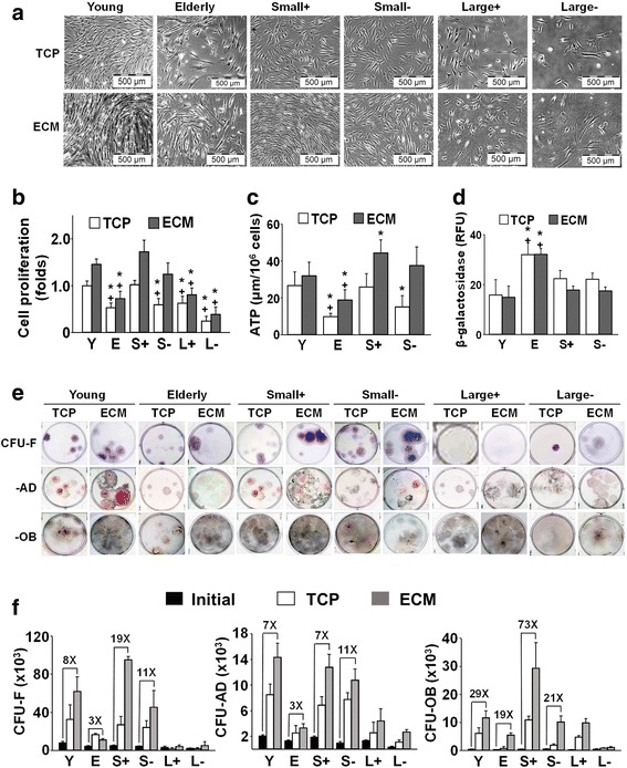
Fig. 4.

Expansion of elderly MSC subpopulations on ECM increases cell number and preserves stemness of small(+) cells. a, b Brightfield microscopy of young and elderly MSCs and isolated subpopulations cultured for 7 days on TCP or ECM revealed that growth on ECM significantly enhanced proliferation of young MSCs and both types of small MSCs. Proliferation was calculated as a fold-change by normalizing cell counts at the end of culture to young cells on TCP (n = 16 donors (11 elderly, five young) tested in replicate experiments). c, d ATP levels, but not β-galactosidase expression, were significantly increased in small(+) and small(–) cells and elderly MSCs with culture on ECM (vs TCP) for 7 days, suggesting that ECM promoted maintenance of cell metabolism and inhibited senescence. There were insufficient numbers of large(+)/large(–) cells for assay (n = 10 donors (five elderly, five young) tested in replicate experiments). e, f Following culture on TCP or ECM for 7 days, young and elderly MSCs and isolated subpopulations were detached and seeded at clonal density on TCP for CFU replication assays (CFU-F, CFU-AD, and CFU-OB). The CFU results were consistent with the proliferation data. Culture of young MSCs on ECM significantly enhanced CFU-AD and CFU-OB production, but not CFU-F. Small(+) cells similarly displayed a significant increase in CFU-AD and CFU-OB, as well as CFU-F, production with culture on ECM. CFU replication was calculated by determining the number of CFUs post culture on ECM or TCP and dividing by the number of CFUs produced by the initial population of cells. Fold increases over the initial number of CFUs are shown (n = 10 donors (five elderly, five young) tested in replicate experiments). *P < 0.05, vs young MSCs; +P < 0.05, vs small(+) MSCs. S small, L large, Y young, E elderly, ECM extracellular matrix, TCP tissue culture plastic, ATP adenosine triphosphate, RFU relative fluorescence units, CFU colony forming unit, F fibroblast, AD adipocyte, OB osteoblast
