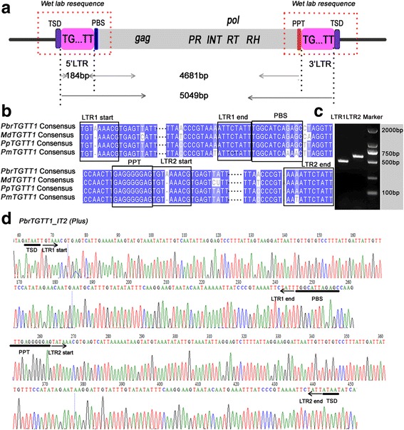
Fig. 1.

Schematic presentation, consensus sequence comparison and wet laboratory verification of TGTT1 elements. a Structural annotations of the TGTT elements. The long terminal repeats (LTRs) are shown in pink boxes; ‘TSD’ indicates the target site duplication; ‘PBS’ indicates the primer binding site; ‘PPT’ indicates the polypurine tract; PR, INT, RT and RH are abbreviations for GAG-pre-integrase, integrase, reverse transcriptase and Ribonuclease H domains, respectively. b The TGTT1 consensus sequence alignment from pear, apple, peach and mei genomes. Identical nucleotides are shown with blue shadows. The internal LTR sequences are marked by ellipsis. c PCR amplification of one randomly selected TGTT1 element (PbrTGTT1_IT2) from the pear genome. The physical positions of the element are located on scaffold809.0 from 128,978 to 133,998. d Resequencing of the PbrTGTT1_IT2 element
