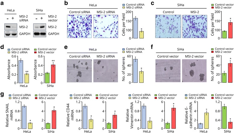
Fig. 2.

MSI-2 overexpression promotes invasion, proliferation and sphere formation of CC cells. a, Western blotting analysis of MSI-2 in indicated cells. GAPDH served as the loading control. b and c, Representative images (left) and quantification (right) of invaded Hela cells (b) and SiHa cells (c), as analyzed using Matrigel invasion assay. d, Cell proliferation was measured using a cell counting kit-8 assay. e and f, Representative images (left) and quantification (right) of sphere formed from HeLa (e) and SiHa (f) cells. g, qRT-PCR analysis of SNAIL, Vimentin, CD44 and E-cadherin in HeLa cells. * P < 0.05
