Fig. 2.

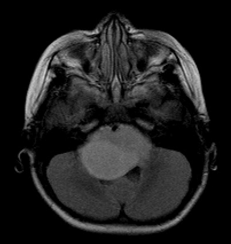
Axial fluid-attenuated inversion recovery image shows a large expansile hyperintense tumor in the pons and brachium pontis with partial encasement of the basilar artery anteriorly and partial effacement of the fourth ventricle posteriorly.

Axial fluid-attenuated inversion recovery image shows a large expansile hyperintense tumor in the pons and brachium pontis with partial encasement of the basilar artery anteriorly and partial effacement of the fourth ventricle posteriorly.