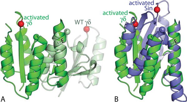
FIGURE 5.

Intramolecular conformational changes upon activation. (A) Superposition, using the E-helices as guides, of one subunit from an activated, DNA-bound γδ resolvase tetramer (green) in a post-cleavage state with one from an inactive wild-type γδ dimer (light green) (PDBids 2gm4 and 2rsl (46, 49)). Red spheres mark the α carbons of the active site serines (S10 for γδ, S9 for Sin); green and blue spheres those of the probable general acid R71 (γδ)/R69 (Sin). (B) Similar superposition of the same activated γδ subunit as in (A), and one subunit from an activated Sin tetramer that appeared to be in the cleavage-ready state (PDBid 3pkz, (50)). doi:10.1128/microbiolspec.MDNA3-0045-2014.f5 49–51). doi:10.1128/microbiolspec.MDNA3-0045-2014.f6
