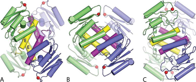
FIGURE 6.

Structures of serine recombinase tetramers in three different rotational states. Colors are as in Figure 4, except that the E-helices are highlighted in yellow (green subunits) and magenta (blue subunits). (A) Activated γδ resolvase tetramer; (B) activated Sin resolvase tetramer; (C) activated Gin invertase tetramer. PDBids 2gm4, 3pkz, and 3uj3, respectively (49–51). doi:10.1128/microbiolspec.MDNA3-0045-2014.f6
