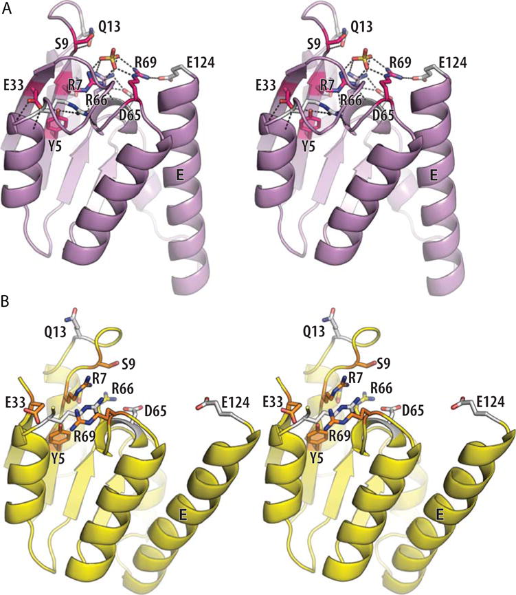
FIGURE 9.

Details of the Sin active site. (A) Stereo view of one subunit from an activated Sin tetramer (PDBid 3pkz (50)). A sulfate ion marks the scissile-phosphate binding pocket, and side chains important for catalysis are shown as sticks. Those residues whose mutation had the most deleterious effect on the rate of DNA cleavage by Tn3 resolvase are shown in magenta, shading to white for those whose mutation had more moderate effects (74). (B) Stereo view of one subunit from a site II-bound wild-type Sin dimer (PDBid 2r0q (64)). The same side chains are shown, similarly shaded from a dark color to white. doi:10.1128/microbiolspec.MDNA3-0045-2014.f9
