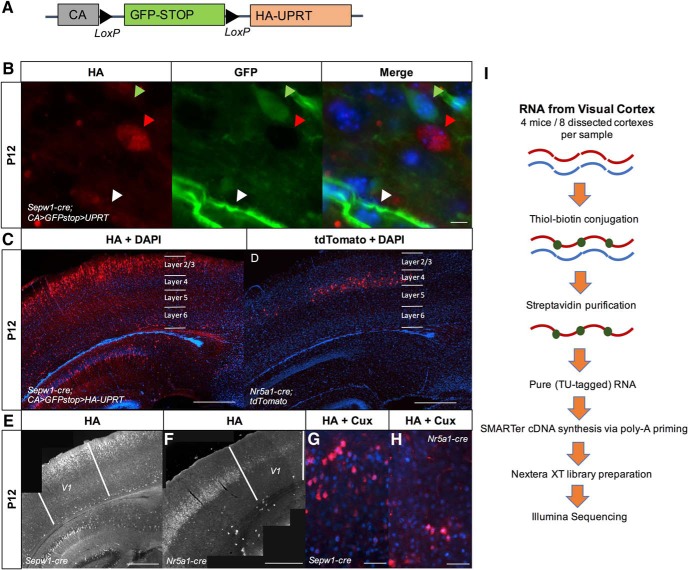
Figure 1.
Upper cortical layer-enriched neuronal expression of HA-UPRT. A, Diagram of the CA>GFPstop>HA-UPRT transgene (Gay et al., 2014). B, Expression of the CA>GFPstop>HA-UPRT transgene (Gay et al., 2013) crossed to a Sepw1-cre line in neurons and endothelial cells (40× objective, scale bar: 10 μm). Green arrow: GFP-positive, Cre-negative neuron; white arrow: GFP-positive, Cre-negative endothelial cell; red arrow: UPRT:HA-positive, Cre-positive neuron. C, Sepw1-cre drives UPRT expression in layer 2/3 and to a lesser extent layer 4. Immunostaining for HA at P12 in a Sepw1-cre; CA>GFPstop>HA-UPRT cross reveals layer 2/3-enriched expression (10× objective, scale bar: 500 μm). DAPI is included to show cortical structure. D, Nr5a1-cre drives expression in a sparse subset of layer 4 neurons. Nr5a1-cre crossed to a tdTomato marker at P12 is specific to layer 4 (10 objective, scale bar: 500 μm). DAPI is included to show cortical structure. E, F, UPRT: HA is expressed in visual cortex of Sepw1-cre (E) or Nr5a1-cre (F) mouse lines crossed to the CA>GFPstop>HA-UPRT line (5× objective, scale bar: 500 μm). G, H, Sepw1-cre and Nr5a1-cre drive expression in neurons found predominantly in layer 2/3 or layer 4 of visual cortex, respectively. Neurons immunostained for the HA epitope on UPRT (red) in Sepw1-cre; CA>GFPstop>HA-UPRT or Nr5a1-cre; CA>GFPstop>HA-UPRT crosses, express the upper layer neuronal marker Cux1 (blue) (40X objective, scale bar: 50 μm). I, TU-tagging workflow using poly-A priming for cDNA synthesis and Nextera XT for library preparation.

