
Figure 2.
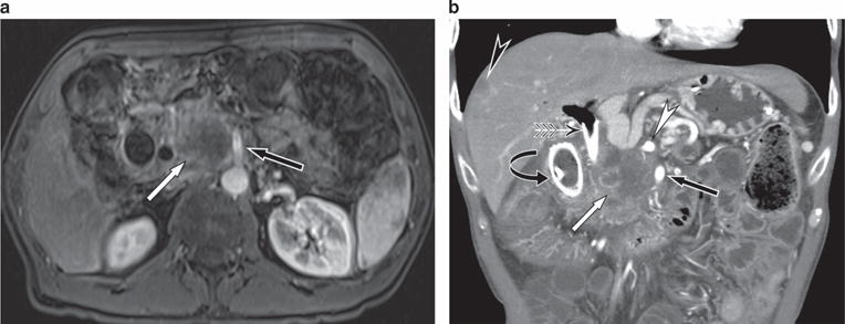
A 53-year-old male presents with painless jaundice. (a) Axial postgadolinium magnetic resonance imaging (MRI) shows a large mass in the pancreatic head (white arrow) closely applied to the superior mesenteric artery (SMA) (black arrow). The tumor was deemed unresectable because of the long length of contact with the SMA. (b) Coronal computed tomography (CT) performed 2 months later shows metal biliary (winged arrow) and duodenal (curved arrow) stents in place. There remains a large tumor (white arrow) that is closely applied to the SMA (black arrow) and celiac artery (white arrowhead). In addition, a liver metastases (black arrowhead) is seen.