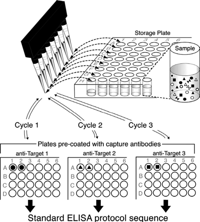
Fig. 3.
A conceptual Illustration of the sequential ELISA. After the samples are subjected to one ELISA for cytokine 1, they are transferred back to a source plate for storage until they are used in the ELISA for cytokine 2, so on and so forth. The steps after the sample incubation are as normal for the particular ELISA adapted from ref. 6.

