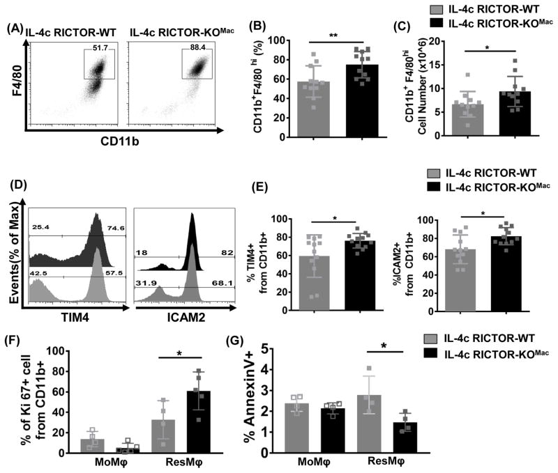
Figure 2. mTORC2 deficiency increases IL-4c tissue-resident peritoneal macrophages via proliferation and cell survival.
RICTOR-WT and RICTOR-KOMac were treated with IL-4c on days 0 and 2. The phenotype of cells from the peritoneal cavity was analyzed by flow cytometry on day 4. (A) Representative flow plot of CD11b and F4/80 and (B) percentages and (C) numbers of CD11b+F4/80hi (N=11/group). (D and E) Representative flow histogram plot and percentages of ResMφ markers (TIM4 and ICAM2) expression gated from CD11b+ population (N=11–12/group). (F) Percentages of proliferation marker Ki-67 gated from CD11b+ population. (G) Percentages of cell death by Annexin V staining on CD11b+TIM4−F4/80lo (MoMφ) and CD11b+TIM4+F4/80hi (ResMφ) Data are compilation of two independent experiments (A–E) or representative of at least three independent experiments (F–G). *P < 0.05, **P < 0.01, Mann-Whitney t tests.

