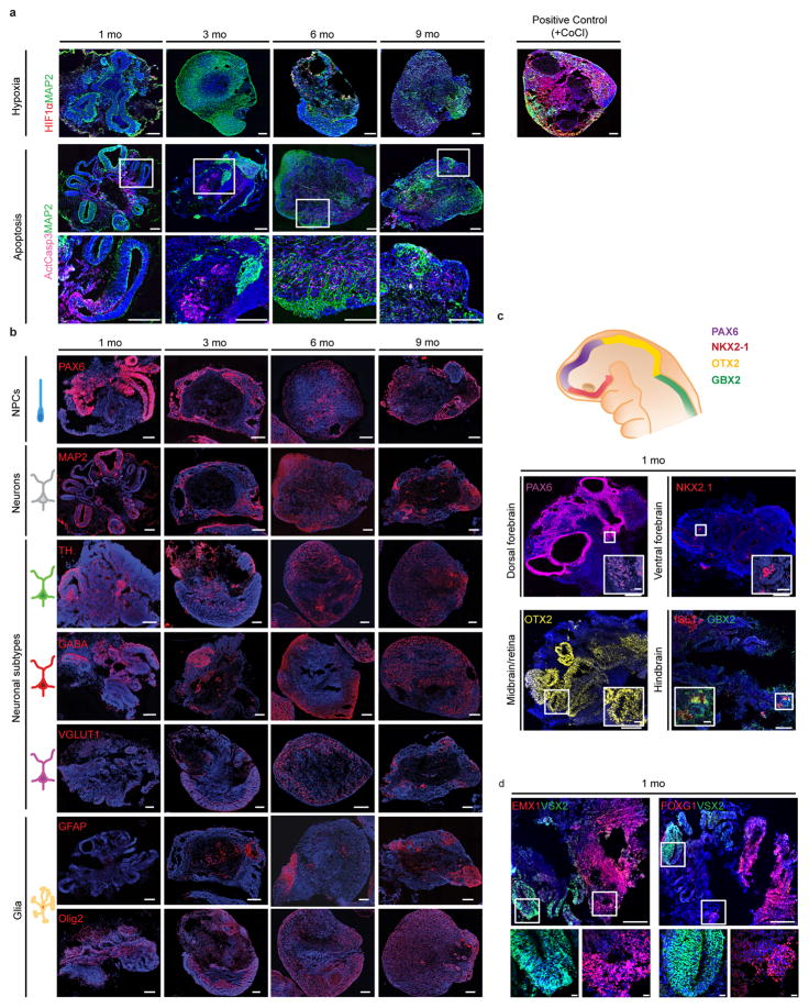
Extended Data Figure 1. Time-course of expression of selected marker genes in human brain organoids.
a. Top: Expression of the hypoxia marker HIF1-α over 1 to 9 months of culture, and positive control (a 6 month organoid treated with cobalt (II) chloride, an activator of the hypoxia signaling pathway). Bottom: Expression of the apoptosis marker active caspase 3 over 1 to 9 months of culture. b. Expression of markers for progenitor, neuronal and glial populations over 1 to 9 months of culture. c. One month old brain organoids exhibit early brain regionalization, expressing markers of forebrain, midbrain, and hindbrain progenitors. d. One month old brain organoids express the cortical marker EMX1, the forebrain marker FOXG1, and the retina marker VSX2, with spatial segregation between regions positive for forebrain versus retinal markers. Scale bars, 250 μm (low magnification), 20 μm (high magnification).

