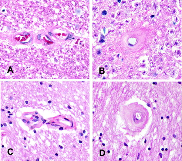
Figure 2. Spinal and Brain Arteriolosclerosis.

Photomicrographs depict the similarity of the arteriolosclerosis grades in the cervical spinal cord and anterior watershed region of the brain. Spinal cord photomicrographs from Figure 1 show no arteriolosclerosis, grade 0 (A) and severe arteriolosclerosis, grade 6 (B). Similar caliber vessels in the brain show no arteriolosclerosis, grade 0 (C) and severe arteriolosclerosis, grade 6 (D). Original magnification × 200.
