Figure 3.
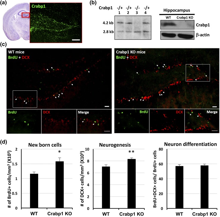
Increased cell proliferation and neurogenesis in the Crabp1 KO mouse hippocampus. (a) Confocal microscopy images showing Crabp1 expression patterns in the hippocampal dentate gyrus. Hematoxylin and eosin image shows a representative dentate gyrus according to the Mouse Brain Atlas second edition. Scale bar = 100 μm. (b) Mouse genotyped by Southern blotting and the lack of Crabp1 expression in the Crabp1 KO hippocampus is validated by Western blotting. (c) BrdU and DCX staining of the dentate gyrus, with magnified images of the boxed areas shown at the bottom. Scale bar = 20 μm. (d) Quantitative analyses showing increases in BrdU+ cells (left, P = 0.02) and DCX+ cells (middle, P = 0.003) in Crabp1 KO, but a similar fraction (∼57%) (right, P = 0.0751) of newborn cells progressed to immature neurons 1 day after BrdU injection in the WT and Crabp1 KO groups (n = 6 per group). Results are presented as mean ± standard error of the mean. *P < 0.05, **P < 0.01 compared with the WT group.

