Figure 2.
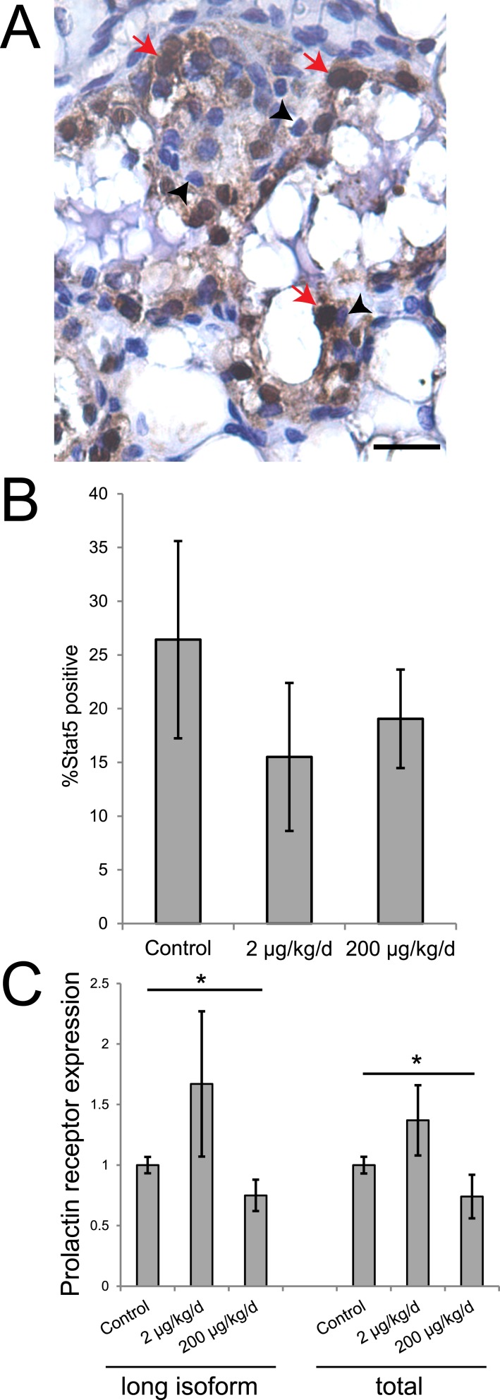
BPS exposure alters expression of prolactin receptor in the mammary gland. (A) A mammary gland sample evaluated for Stat5 expression. Scale bar represents 20 μm; red arrows indicate positive cells; and black arrowheads indicate negative cells. (B) Quantification of the percentage of mammary epithelial cells expressing Stat5 on LD21. (C) qRT-PCR reveals significant differences in the expression of prolactin receptor (long isoform and total) between females from the three treatment groups. *P < 0.05, independent samples median test, post hoc not available. For qRT-PCR, sample sizes were as follows: control (n = 5), 2 μg BPS/kg/d (n = 6), 200 μg BPS/kg/d (n = 6). For immunohistochemistry, sample sizes were as follows: control (n = 9), 2 μg BPS/kg/d (n = 11), 200 μg BPS/kg/d (n = 10).

