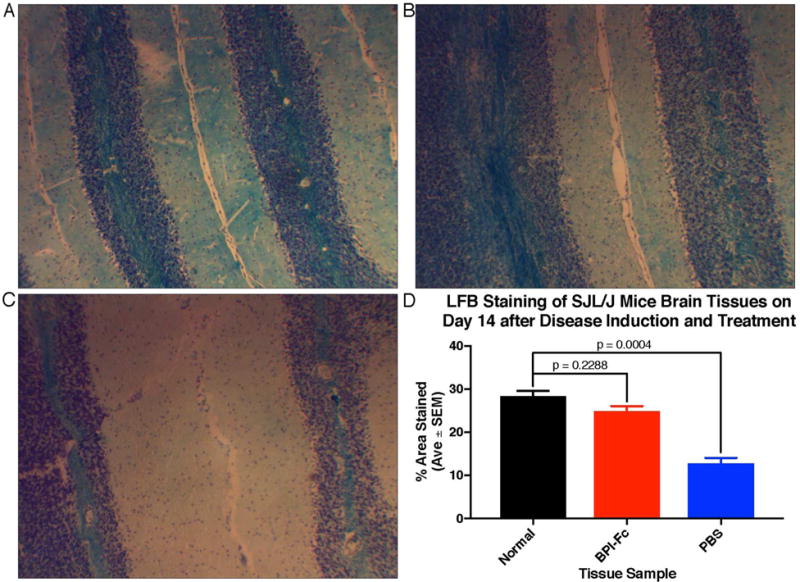
Figure 7.

Luxol fast blue staining of mice brain slices on day 14 after EAE induction. (A) Normal: mouse neither induced with EAE nor treated. (B) BPI-Fc: mouse induced with EAE and treated with BPI-Fc. (C) PBS: mouse induced with EAE and treated with PBS. (D) Quantification of the area stained by LFB in each tissue. The % Area Stained by LFB for each tissue was measured in triplicate, and the results between the three tissue samples were compared for statistical significance using a one-way ANOVA and Tukey multiple comparisons test.
