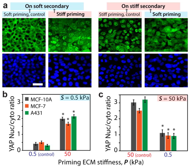
Figure 6. YAP activity depends on past ECM stiffness.

(a) Immunofluorescent staining of MCF10A cells for YAP (green) and DAPI (blue) illustrating the subcellular localization of YAP for the monolayer migrating on secondary ECM, after priming. Scale bar = 50 μm. (b,c) Average nuclear-to-cytoplasmic ratio of the YAP fluorescent intensity for MCF10A, MCF7, and A431 cells within the monolayer. *p3<30.05 with respect to control ECMs of homogeneous stiffness. Error bars = SEM. N>40.
