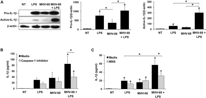
Figure 2. Viral infection augments LPS-induced FM IL-1β processing and secretion through activation of the NLRP3 inflammasome.
(A) Human FM explants were treated with (A) No treatment (NT), LPS (100ng/ml), MHV-68 (1.5×10⁴/ml PFU) or both MHV-68 and LPS (n=7). Lysates were evaluated by Western blot for expression of pro-IL-1β (31kDa) and the 17kDa active form. Blots are from one representative experiment. Bar charts show pro- and active-IL-1β expression as determined by densitometry and normalized to β-actin (n=4–5). (B & C) Human FM explants were treated with NT, LPS, MHV-68 or both MHV-68 and LPS in the presence of either media or (B) a caspase-1 inhibitor (1µM) (n=6); or (C) the NLRP3 inhibitor, MNS (10µM) (n=5). Supernatants were measured for IL-1β by ELISA. *p<0.05 relative to the NT control unless otherwise indicated. Data are expressed as mean±SEM.

