
Fig. 1. Inhibition of sEH or genetic deletion decreases diclofenac-induced ulcers in stomachs of mice.
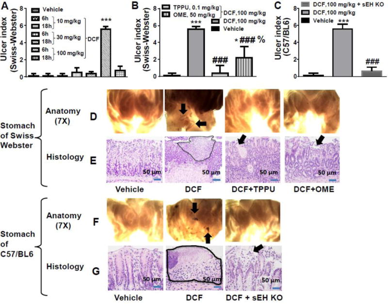
(A) Among the 3 doses tested for the ulcerative effect of diclofenac (DCF), the 100 mg/kg dose exhibited ulcers in Swiss Webster mice (*** p < 0.001 vs 10 mg/kg dose). The severity of the ulcers was greater at 6 h post diclofenac dose than at 18 h after diclofenac treatment. (B) 100 mg/kg of diclofenac-induced stomach ulcers significantly in Swiss Webster mice (*** p < 0.001, DCF treated animals vs vehicle control animals; * p < 0.01, DCF + OME vs vehicle-treated animals). Severity of diclofenac-induced ulcers decreased significantly with TPPU or OME treatment (### p < 0.001, DCF vs DCF + TPPU, DCF vs DCF + OME). OME-treated animals exposed to DCF had higher (% p < 0.01) incidence of ulceration than TPPU + DCF treated animals. (C) DCF significantly induced stomach ulcers in C57BL6 mice (*** p < 0.001, DCF vs vehicle). The sEH gene deletion (KO) also decreased ulcerative effects of diclofenac in stomach (### p < 0.001). The vehicle control animals used for this figure are C57BL6 mice. Values are presented as the mean ± standard deviation, n=4. (D) Anatomical observation at 7X under a dissecting microscope of stomachs of Swiss Webster mice administered with different treatments. Ulceration is pointed with an arrow. Stomachs from mice exposed to DCF exhibited ulcers and hemorrhagic streaks. Pre-treatment with the sEHI TPPU, omeprazole (OME), or (F) sEH knock out (KO) decreased stomach ulceration as shown at 7X under a dissecting microscope. (E) Histopathological observation of tissue section complemented anatomical observation. Morphology of gastric mucosa of vehicle treated Swiss Webster mice looked normal while ulceration was observed on stomachs of DCF treated mice. Luminal surface necrosis tissue debris is shown as highlighted area and it’s beneath granulation tissue formation is noticed. Ulceration was minimal in mice treated with DCF + TPPU. Minimal glandular cystic injury/erosion on surface of the mucosal layer in TPPU pre-treated mice is shown with an arrow. Omeprazole also minimized gastric erosion due to DCF. Minimal erosion on surface of mucosal layer is pointed by arrow. (G) Gastric mucosa of C57BL6 mice was normal but ulceration in mucosa (highlighted area) was observed in DCF treated mice. Mucosa with minimal injury/erosion and predominant reactive epithelial changes is seen in stomachs of sEH knockout mice. Doses of TPPU and OME were 0.1 mg/kg and 50 mg/kg respectively.