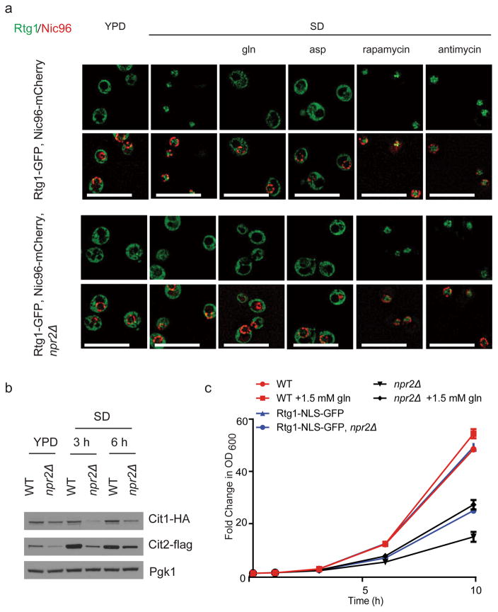
Figure 3. npr2Δ mutants exhibit a defective mitochondria-to-nucleus retrograde (RTG) response.
(a) WT or npr2Δ cells expressing Rtg1-GFP, Nic96-mCherry (nuclear pore complex marker) were transferred from YPD to SD for 6 hours and treated with mock, 50 μM antimycin, 50 nM rapamycin, 2 mM glutamine or 2 mM aspartate (pH 5.1) for 30 min prior to imaging. Antimycin and rapamycin treatment activated the retrograde response by in both strains, while glutamine or aspartate addition inactivated the pathway in WT cells. Scale bar 5 μm.
(b) Western blot depicting amounts of retrograde response targets Cit1p and Cit2p in WT and npr2Δ cells switched from YPD to SD for indicated times. Note that npr2Δ mutants have consistently lower amounts of these enzymes in SD medium. Full gels are shown in Supplementary Fig. 5c.
(c) Forced nuclear localization of Rtg1p can partially rescue the growth of npr2Δ mutants. Growth of WT or npr2Δ cells expressing a version of Rtg1-GFP containing a strong nuclear localization signal (NLS) (Rtg1-NLS-GFP). Two duplicates at each time point were measured, in two independent experiments.

