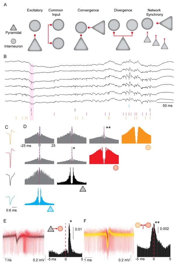
Figure 1. Synaptic interactions, common drive and circuit motifs inferred from spike train correlations.
A. Circuit motifs hypothesized to result in short-latency spike-spike correlations. B. Example wideband (0.1– 6,000 Hz) extracellular traces obtained from dorsal CA1 pyramidal layer. Colored ticks represent spikes from single units sorted offline. Pink shaded area is a putative instance of monosynaptic spike transmission from a pyramidal neuron (black tick) to an interneuron (red tick). C. Mean waveforms for the four units shown in B. D. Autocorrelations (in color) and CCGs (in grey) for the four units from B and C. Dashed line shows 0 ms lag from the reference spike. CCG binned at 1 ms. Note that both pyramidal neurons have positive (~ 1 ms) latency peaks in their pairwise CCGs with the interneurons (*), while the CCG between the two interneurons has a peak at 0 ms lag (**). E. Left: 4,000 randomly sampled traces (filtered at 0.3 – 6 kHz) aligned to the spike of the pyramidal neuron (in black), with the spikes from the postsynaptic interneuron (in red), from the pair highlighted with the asterisk in D. Right: High resolution CCG (0.1 ms bins) for the same pair. Vertical scale bar is non-corrected probability. F. Left: 4,000 randomly sampled traces (filtered at 0.3 – 6 kHz), aligned to the spike of the interneuron in yellow, with the spikes from the second interneuron in red, from the pair highlighted with the double asterisk in Panel D. Right: High resolution CCG (0.1 ms bins) for the same pair. Vertical scale bar is non-corrected probability.

