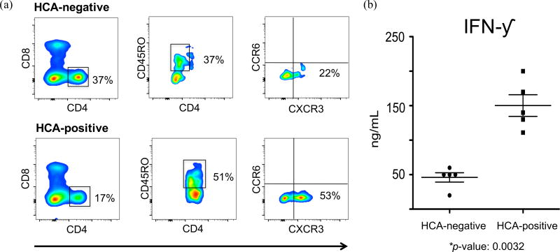
Figure 6. Exposure to HCA increases the percentage of memory CD4+ T lymphocytes exhibiting a TH1 phenotype and IFN-γ secretion.
(a) Representative analytic flow cytometry plot of CD4+ T lymphocytes collected on postnatal day 10 from 5 pairs of preterm infants matched for gestational age, day of blood draw, sex, race, prenatal steroid exposure, and delivery mode. Each pair consisted of one HCA-positive and one HCA-negative patient (10 samples total). Here we are showing data from two 24 weeks gestation female Hispanic infants, delivered via Cesarean section following prenatal steroid prophylaxis. One infant was exposed to in utero HCA while the other showed no clinical or histological signs of chorioamnionitis.
(b) IFN-γ secretions were measured by Luminex technology. Cells were collected from 5 pairs of preterm infants matched for gestational age, day of blood draw, sex, race, prenatal steroid exposure, and delivery mode. Each pair consisted of one HCA-positive and one HCA-negative patient (10 samples total). Paired t-test was used to determine statistical significant differences. This experiment was independently performed once, without replicates.

