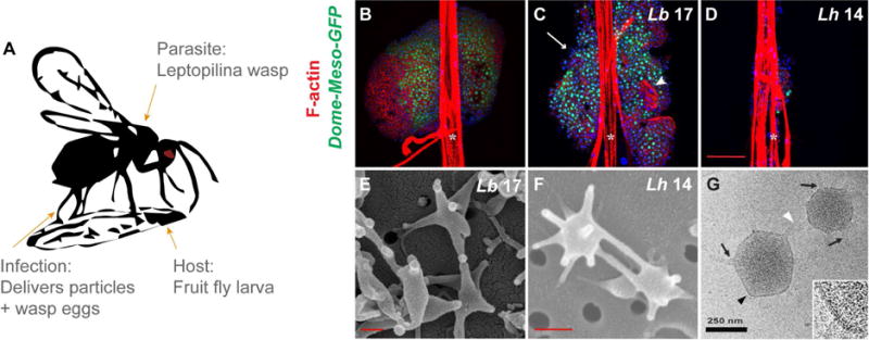
Fig 1. Effects of wasp attack on host lymph glands and comparison of VLP morphologies.

(A) Infection by female Leptopilina spp. parasitic wasps introduces not only wasp eggs into the body cavities and hemolymph of fruit fly larvae, but also venom gland products which includes spiked, 300-nm VLPs. VLP bioactivity is known to be necessary for the infective success of L. heterotoma, rather than other venom constituents [6, 9]. (B) Intact anterior lymph gland lobes from uninfected control Dome-MESO-GFP fly larvae. GFP marks the stem-like progenitors in the medulla. (C) Dome-MESO-GFP glands of Lb 17-infected larvae show lamellocyte differentiation (white arrowhead) and lobe dispersal (white arrow). (D) Progenitors are depleted in Dome-MESO-GFP anterior lobes infected with Lh 14. (B – D) White asterisks mark dorsal vessels. (E) Scanning EMs of Lb 17 and (F) Lh 14 VLPs. (G) CryoEM of Lh 14 VLPs: The external lipid bilayer is contiguous, extending from spike bases (black arrows) at the VLP core to spike tips (white arrowhead). The black arrowhead marks area in zoom, bottom right.
