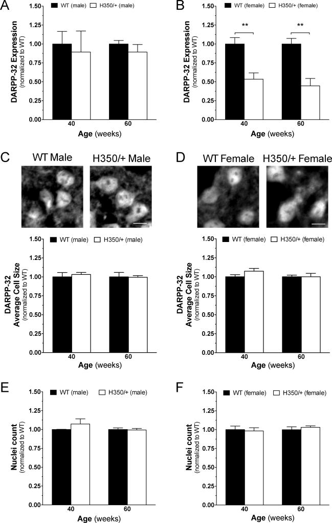
Figure 8. HdhQ350/+ females, but not males, exhibit typical HD protein loss in the striatum.
(A) Male HdhQ350/+ mice did not show changes in DARPP-32 protein expression at 40 or 60 weeks when compared to male wild-type littermates. (B) Female HdhQ350/+ mice showed significant reductions in DARPP-32 protein expression at 40 and 60 weeks of age. (C) Male HdhQ350/+ and (D) female HdhQ350/+ mice had no significant differences in average size of DARPP-32-positive cells compared to respective wild-type littermates. Representative images are cropped and captured at 40×. Scale bar denotes 10um. DAPI-positive cells quantified in the dorsolateral striatum showed no difference in (E) male HdhQ350/+ and (F) female HdhQ350/+ mice when compared to their respective wild-type littermates. N=3 for all groups. Error bars represent S.E.M. and **p<0.01, with two-way ANOVA with Bonferroni post-hoc test.

