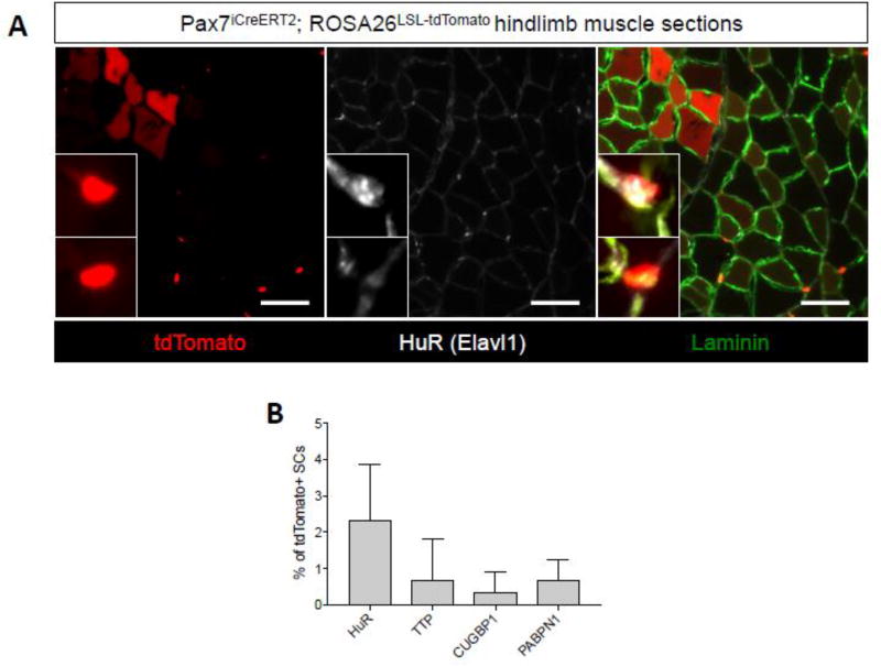
Figure 7.
Heterogeneous RNA binding protein expression in muscle SCs. (A) Representative immunofluorescence images of TA muscle tissue sections from tamoxifen treated Pax7iCreERT2;ROSA26LSL-tdTomato mice stained with an antibody targeting HuR (white). Laminin is shown in green, nuclei are labeled with DAPI (blue) and tdTomato fluorescence is shown in red. Insets are of a tdTomato+::HuR+ cell (top inset) and a tdTomato+::HuR- cell (bottom inset) (B) A graph quantifying the percent positive cells for each indicated RNABP as a fraction of tdTomato+ SCs. Error bars represent SD from n=3 mice.

