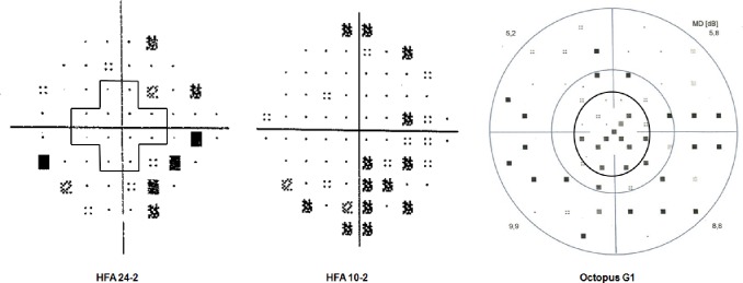
Fig 1. Pattern deviation plots of the Humphrey Field Analyzer (HFA) 24–2 test and the HFA 10–2 test, and the corrected probability plot of the Octopus G1 program of the same early glaucoma eye from the study.
The pattern deviation plot of the HFA 24–2 test shows no depressed test point within the central 10° area (the area surrounded by the cross shape line) but the sensitivity is reduced in the more peripheral area of the visual field. The pattern deviation plot of the HFA 10–2 test shows a cluster of depressed test point locations. The peripheral retinal sensitivity is not tested. The corrected probability plot of the Octopus G1 program shows a cluster of reduced sensitivity both in the central 10° area (the region within the inner black circle) and in the periphery.

