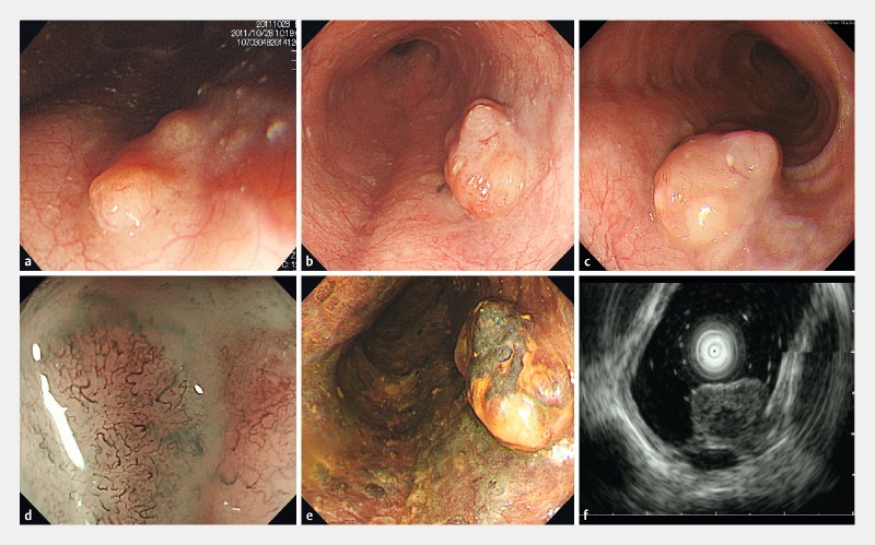
Fig. 1.

Endoscopic examination. a Esophageal SMT at first detection. b, c A well-defined non-pigmented elevated lesion with a flat proximal edge 2 years after first detection. d Narrow band imaging magnification endoscopy showing subepithelial structures resembling reticular vessels. e Iodine staining. f Endoscopic ultrasonography depicted the tumor as a well-circumscribed low-echoic mass within the submucosal layer.
