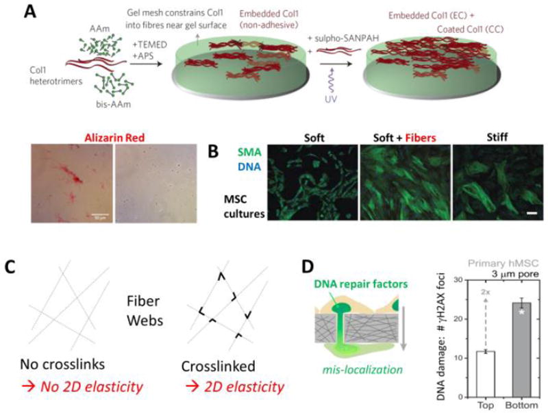
Figure 4. Fibrous composites, fiber crosslinking, and rigid pore effects on DNA damage in migration.
A. Scar-like islands of collagen-I are heterogeneously entrapped at the subsurface of the soft hydrogel in order to generate minimal matrix models of scars, MMMS. AAm (acrylamide) polymerizes and is crosslinked by bis-AAm when initiated and catalyzed by TEMED and APS; sulfo-SANPAH and UV allow uniform attachment or coating of collagen on the gel surface. Heterogeneity of the MMMS was confirmed by both immunofluorescence and staining with the histochemical dye widely used to visualize fibrosis, Sirius Red. Adapted from [47].
B. Human MSCs cultured on conventional soft or stiff homogeneous gels and separately on MMMS, which is soft + fibers; on the latter two substrates, the MSCs upregulate a common cell marker of cell tension and fibrosis, smooth muscle actin (SMA). Results are adapted from [47].
C. A web if fibers that is not crosslinked has no lateral stiffness or 2D elasticity, but addition of crosslinks or welds at fiber junctions will generate a non-zero 2D elasticity.
D. MSC migration through small pores can rupture the nuclear envelope and exhibit an increase in DNA damage measured in terms of immunostained foci of phosphorylated histone H2AX (i.e. γH2AX). Results are adapted from [54].

