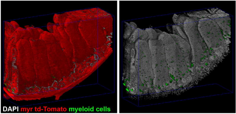
Figure 2. Tissue clearing and imaging of mesoscale 3D specimen generates complex, high resolution volumes.

Intestine from FLT3Cre+; ROSA26mTmG/mTmG mice expressing myristoylated-td-Tomato(myr td-Tomato, red) and GFP in myeloid cells(myeloid cells, green) were dissected, stained for nuclei(DAPI, gray) and cleared with Triton X-100 in phosphate buffered saline. Multiple overlapping volumes were imaged by confocal laser scanning microscopy and mosaic merged. At left, DNA and GFP are shown to demonstrate the number and complexity of the enterocytes and intercalated myeloid cells.
