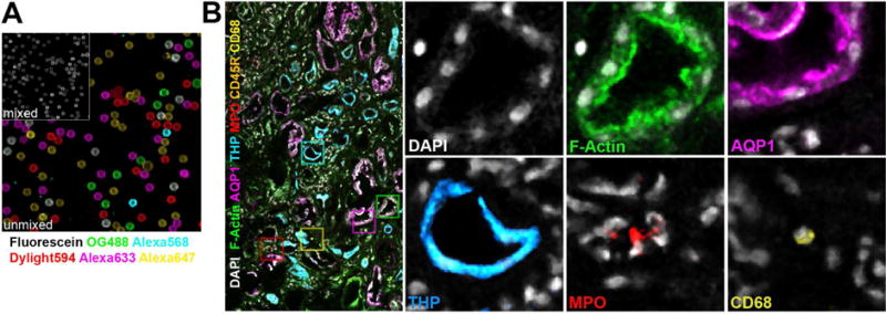
Figure 3. Spectral unmixing of seven labels in a human kidney biopsy volume.

Beads and the biopsy were labeled with seven fluorophores. Multiple overlapping confocal volumes of the human biopsy were collected and mosaic merged. Spectra from the independently labeled beads were used to spectrally unmix the seven fluorophores with LASX(Leica). (A) Secondary antibodies independently absorbed to latex beads were combined and then spectrally unmixed with reference spectra. Reference spectra were collected from individually labeled beads. (B) Human biopsies were stained for nuclei (DAPI, gray), F-actin (phalloidin-Oregon Green 488, green) and species specific secondary antibodies labeled with Dylight594, Alexa647, Fluorescein, Alexa633 and Alexa568 were used to label primary antibodies against myeloperoxidase (MPO, red), macrophages (CD68, yellow), B-cells (CD45R, orange), Aquaporin-1(AQP1, magenta) and Tamm-Horsfall Protein (THP, cyan).
