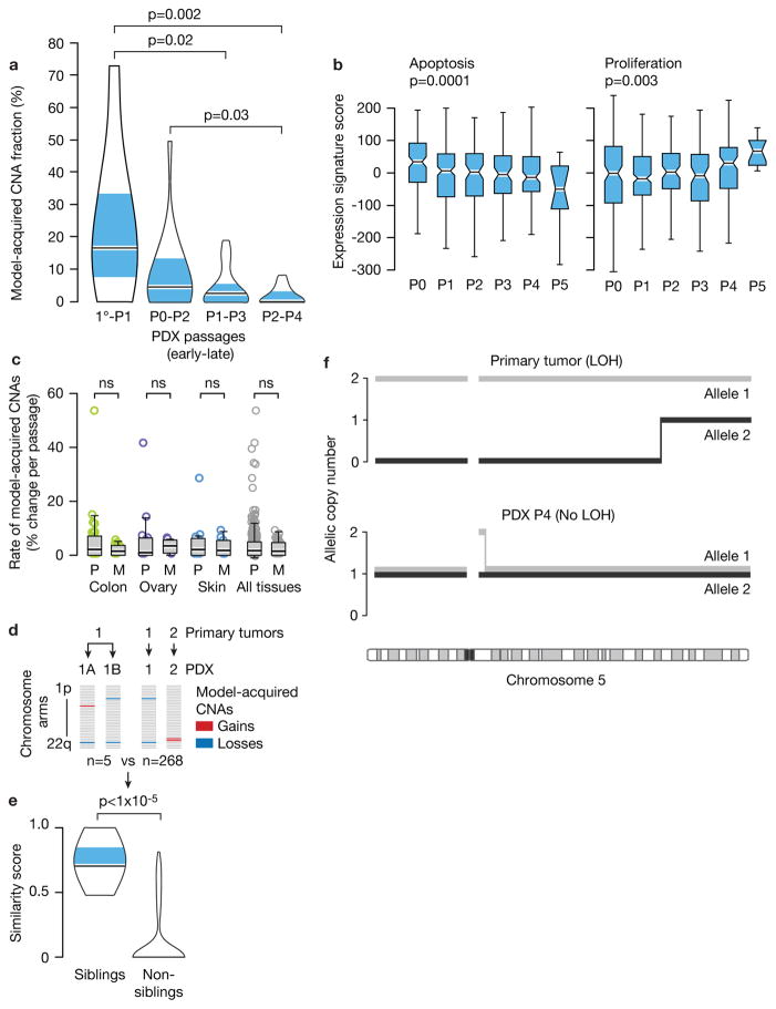
Figure 2. Selection of pre-existing subclones underlies CNA dynamics.
(a). The rate of model-acquired CNAs decreases with PDX passaging. Violin plots present the fraction of CNAs acquired within two in vivo passages as a function of passage number. P-value indicates significance from a Wilcoxon rank-sum test. 1°, primary tumor. (b) Apoptosis decreases and proliferation increases with PDX passaging. Box plots present the apoptosis (left panel) and proliferation (right panel) gene expression signature scores as a function of passage number. P-values indicate significance from a Kruskal-Wallis test. (c) Similar CNA acquisition rates in PDXs from primary tumors and from metastases. Box plots present the rate of model-acquired CNAs as a function of tumor source (P=primary, M=metastasis), across three available tissue types. n.s., non-significant (Wilcoxon rank-sum test). (d) Schematics showing the calculation of pair-wise similarity scores for PDX models coming from the same primary tumor but propagated independently in the mouse (“sibling” PDXs; n=5) and for PDX models coming from distinct primary tumors (“non-sibling” PDXs; n=268). (e) “Sibling” PDXs tend to acquire more similar aberrations than lineage-matched “non-sibling” PDXs. Violin plots present the similarity scores of “sibling” and “non-sibling” PDXs. P-value indicates significance from a lineage-controlled permutation test. (f) Alleles that seem to have been lost in primary tumors can “re-appear” in PDXs, demonstrating expansion of rare pre-existing subclones throughout PDX propagation. Plots present the copy number of both of chromosome 5 alleles in a primary tumor and its derived PDX. Loss of heterozygosity (LOH) is identified in the primary tumor along most of chromosome 5, but both alleles are detected in a 1:1 ratio in the PDX derived from that primary tumor.

