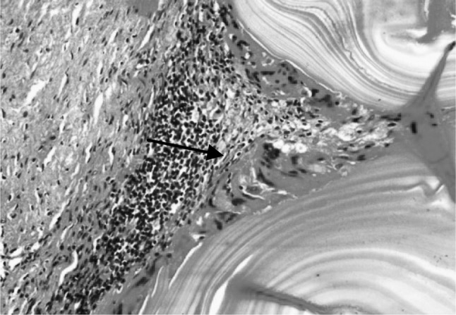
Fig. 7.

Section of the sheep liver affected with hydatidosis revealing laminated cyst wall surrounded immediately by macrophage cell layer (arrow) followed by a layer of infiltrating cells of eosinophils and mononuclear cells and fibroblastic cell layer. H&E–Original magnification ×400X
