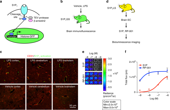
Fig. 6.
S1P1 activation by LPS in the brain. a Schematic of the design to monitor S1P1 activation in S1P1 GFP signaling mice. S1P1 is fused to a transcription factor (TF) via a protease recognition sequence. Ligand binding to the receptor stimulates the recruitment of a β-arrestin2–TEV protease fusion protein, triggering the release of the transcription factor from the C terminus of modified S1P1. The transcription factor stimulates expression of a histone-GFP reporter gene, fluorescently labeling cell nuclei. b LPS or vehicle was injected intraperitoneally into S1P1 GFP signaling (S1P1GS) mice, and the brains were harvested 7 days after injection and examined by immunofluorescence. The tissues of three mice injected with LPS and three mice injected with vehicle were examined. c Representative histological sections were immunostained with antibodies to CD31, and the images of cerebral cortex, cerebellum, and brainstem were captured using an inverted laser scanning confocal microscope. Scale bar, 50 μm. d Brain endothelial cells (EC) were isolated from S1P1 luciferase signaling (S1P1LS) mice, grown in triplicate in 24-well plates, treated with S1P or RP-001 and D-luciferin, and immediately subjected to BLI. e Bioluminescence images of the cells exposed to the indicated concentrations of S1P or RP-001 represent a 15-min acquisition. The experiment was repeated twice. f The bioluminescence activity was quantified by determining the total flux (photons/sec; p/sec). Data represent the mean ± SEM, n = 6

