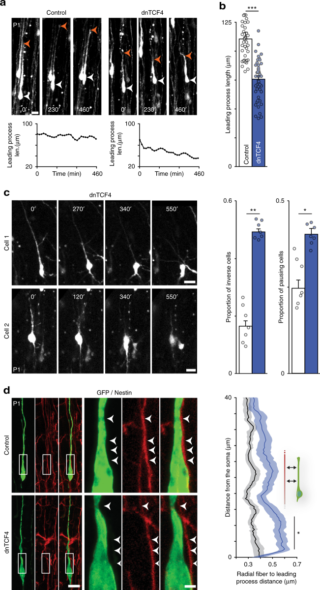
Fig. 2.
Loss of Wnt canonical signaling alters leading process stability and CPN attachment to radial glia. a Confocal time lapse sequence of representative control and dnTCF4 electroporated cells migrating in the cortical plate (CP). Graphs show dynamics of leading process length over time measured in the cells depicted. b Post hoc analysis of confocal images indicates that the leading process of dnTCF4 cells appears shorter than that of controls; n = 50 cells/condition from 7 brains, Student’s t test. See also Supplementary Movie 3. c Confocal time lapse sequences of two representative dnTCF4 cells migrating in the CP showing the frequently observable event of leading process inversion. Graphs represent the percentage of tracked cells exhibiting leading process inversion at least once (inverse cells) and the percentage of tracked cells remaining stationary for at least 100 min (pausing cells); n = 7 brains, Mann–Whitney. See also Supplementary Movie 4. d Representative images illustrating migrating cells attached to nestin-positive radial glia fibers. Note the increased space between the leading process of dnTCF4 electroporated cell and the glia fiber (arrowheads). We performed point-by-point measurement of the distance between the leading process of migrating cells and the radial fiber; n = 50 cells/condition from 4 brains, two-way ANOVA. Graphs display mean ± s.e.m. *P < 0.05, **P < 0.01, ***P < 0.001. Bars = 2 µm (d, close up) and 20 µm (a, c, d)

