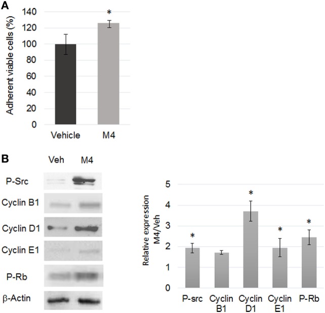
Figure 1.

M4 stimulating cell proliferation. (A) Quantification of MCF-10A cell viability by crystal violet assay after 24 h vehicle or 1 nM M4 exposure. M4 treatment triggered a 26% increase of cell proliferation. Each bar represents mean ± SD. N = 3. *p < 0.05. (B) Western blot analyses of protein expression level (left) and corresponding quantifications (right) of phospho-Src (tyr416), Cyclin B1, D1, E1 and phospho-Rb (retinoblastoma protein) in 24 h vehicle or 1 nM M4-treated cells. β-Actin was used as a loading control. Enhanced Src and Rb phosphorylation as well as Cyclin D1 and Cyclin E1 expression was observed under M4 treatment. Each bar represents mean ± SD. N = 4. *p < 0.05.
