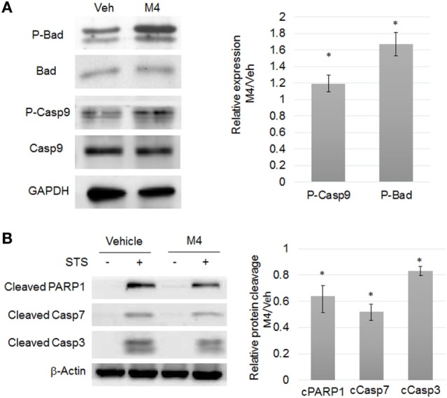
Figure 2.

M4 stimulating resistance to apoptosis. (A) Western blot analyses of protein expression level (left) and corresponding quantifications (right) of phospho-Bad (ser136), phospho-Caspase 9 (ser196) in 24 h vehicle or 1 nM M4-treated cells. M4 treatment triggered a significant 19% and 67% increase of Bad (ser136) and Caspase 9 (ser196) phosphorylation, respectively. Each bar represents mean ± SD. N = 3. *p < 0.05. (B) MCF-10A cells were pretreated for 24 h with vehicle or 1 nM M4, and then exposed to 0.25 µM staurosporine (STS) for 6 h or not (Veh). Cleavage of PARP1, Caspase 7, and Caspase 3 were evaluated with specific antibodies. β-Actin was used as a loading control. Results are represented as M4 + STS/STS ratio. M4 treatment triggered a significant 36, 48, and 17% decrease of PARP1, Caspase 7, and Caspase 3 cleavage, respectively. Each bar represents mean ± SD. N = 4. *p < 0.05.
